




Preparing accurate molar stock solutions of synthetic multi receptor peptides for quantitative in vitro bioassays requires rigorous stoichiometric calculations before any solvent contacts the lyophilized cake. How to Prepare Retatrutide Solutions for Laboratory Evaluation? details the analytical chemistry protocols governing gravimetric weighing, ionic strength adjustment, sterile filtration, and spectrophotometric verification. A foundational rule in analytical peptide preparation is recognizing that gross powder mass never equals active peptide mass. Lyophilized synthetic peptides exist as electrostatic complexes containing bound counter ions, such as acetate or trifluoroacetate, alongside residual bound water molecules. Before weighing, sealed vials removed from minus 80 degrees Celsius storage must equilibrate inside a desiccator to ambient laboratory temperature for at least thirty minutes to prevent atmospheric moisture condensation onto the hygroscopic powder. Using a calibrated microbalance equipped with an antistatic ionizer, analysts calculate true molarity by multiplying the gravimetric weight by both the chromatographic purity percentage and the batch specific net peptide content derived from quantitative amino acid analysis, dividing the resulting active mass by the theoretical molecular weight of 4731.33 Daltons.
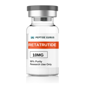
Selecting an appropriate aqueous vehicle for initial dissolution requires balancing the amphipathic surface charge of the 39 amino acid backbone with the hydrophobicity of its C20 eicosanedioic acid conjugate. The theoretical isoelectric point of this chimeric sequence lies in the mildly acidic range near pH 4.5 to 5.5. Attempting to dissolve the lyophilized cake in acidic buffers near its isoelectric point neutralizes net electrostatic surface charge, collapsing the repulsive double layer and triggering immediate isoelectric precipitation. Conversely, reconstituting in high salt buffers with excessive ionic strength compresses the Debye screening length around the peptide monomers, promoting hydrophobic self association into inactive fibrils. Optimal dissolution protocols initiate hydration using sterile, degassed ultra pure water or a weak 10 millimolar ammonium bicarbonate buffer adjusted to pH 7.4 to 7.8. At this slightly alkaline pH, both the glutamate residues on the peptide backbone and the terminal carboxylate of the C20 diacid side chain are fully deprotonated, generating electrostatic repulsion that drives rapid, complete monomer solvation without mechanical vortexing.
When preparing peptide solutions for long term three dimensional organoid cultures or multi day cellular respiration assays, sterile filtration is frequently required to eliminate microbial contamination. However, passing diluted, lipidated peptides through inappropriate syringe filter membranes represents a major source of unquantified compound loss. Nitrocellulose, nylon, and mixed cellulose ester membranes exhibit strong hydrophobic and electrostatic interactions that strip acylated peptides from the filtrate. Analytical recovery evaluations demonstrate that only hydrophilic polyvinylidene fluoride or surface modified polyethersulfone membranes with a 0.22 micrometer pore size provide acceptable recovery rates exceeding 95 percent. To prevent concentration depletion in small volume preparations, analysts must select low hold up volume syringe filters measuring 4 millimeters or 13 millimeters in diameter and pre saturate the membrane matrix with dilution buffer containing 0.1 percent bovine serum albumin or 0.01 percent polysorbate 20 before passing the peptide stock through the housing.
Rather than relying solely on initial gravimetric calculations, rigorous analytical laboratories verify the exact molar concentration of reconstituted stock solutions prior to serial dilution using ultraviolet absorption spectrophotometry. Because the primary sequence of this triple agonist contains two tyrosine residues at positions one and ten, along with aromatic electronic transitions along the peptide backbone, clarified stock solutions exhibit a distinct ultraviolet absorption peak at 280 nanometers. Utilizing a microvolume spectrophotometer or a quartz cuvette with a 1 centimeter pathlength, analysts measure baseline corrected absorbance at 280 nanometers against a matched buffer blank. Checking absorbance at 320 nanometers simultaneously serves as a turbidity quality control check; any measurable reading at 320 nanometers indicates light scattering caused by undissolved micro aggregates. By applying the Beer Lambert law using the sequence specific molar extinction coefficient calculated from its aromatic residue composition, laboratory technicians confirm the exact micromolar concentration in solution before committing the reagent to high throughput screening plates.
Transferring verified stock solutions into 96 well or 384 well assay plates introduces fluid dynamic variables that must be controlled during serial dilution preparation. Due to the C20 fatty diacid conjugate, concentrated peptide solutions exhibit slightly altered surface tension and viscosity compared to pure aqueous buffers, which can cause volume dispensing inaccuracies in air displacement robotic pipettes. Automated liquid handling workstations must be calibrated using liquid class profiles optimized for surfactant containing protein solutions, incorporating slower aspiration speeds and controlled blowout delays. Furthermore, serial dilution matrices must be executed exclusively within certified low binding fluoropolymer or cyclic olefin copolymer microplates. Carrying pipette tips across dilution steps must be strictly prohibited, as peptide molecules adsorbed to the outer polypropylene tip wall during stock aspiration will leach into subsequent low concentration wells, skewing half maximal effective concentration curves by several orders of magnitude.
The precision of any laboratory solution preparation protocol depends ultimately on the chemical homogeneity and analytical documentation of the starting lyophilized material. In accordance with United States Food and Drug Administration bioanalytical quality standards, every stock solution log must link directly to a batch specific Certificate of Analysis confirming chromatographic purity above 98.0 percent, verified mass spectra, and explicit net peptide content percentages. Sourcing synthetic peptides from suppliers operating under certified ISO quality management systems and World Health Organization Good Manufacturing Practice standards eliminates the risk of insoluble synthesis resins or unremoved protecting groups ruining solution clarity. PeptideGurus supports global analytical and pharmacological laboratories by delivering authenticated multi receptor peptides validated through dual third party testing. Backed by temperature monitored cold chain logistics across North America and Europe, research teams receive highly soluble, pristine lyophilized cakes that reconstitute predictably across demanding laboratory evaluations.
Technical Overview Note: The preceding content represents a technical overview and synthesis of industry wide knowledge in peptide science and analytical pharmacology for educational and research reference, rather than conclusions drawn from a single dedicated clinical study.
Why does measuring UV absorbance at 320 nm matter when verifying peptide stock solutions?
Peptides and aromatic amino acids do not absorb ultraviolet light at 320 nanometers. Therefore, any apparent absorbance detected at 320 nm is actually caused by light scattering from suspended sub visible particulates or self assembled peptide aggregates. Monitoring the 320 nm wavelength provides an immediate, non destructive optical quality check to confirm complete monomeric dissolution before using the stock in cellular assays.
Which syringe filter membrane materials cause the highest loss of lipidated peptides?
Nylon and nitrocellulose membranes cause the highest adsorptive loss of lipidated peptides due to strong hydrophobic and hydrogen bonding interactions with the C20 fatty acid chain and peptide amide backbone. To prevent stripping active compound from solution during sterile filtration, laboratory protocols must utilize low protein binding hydrophilic polyvinylidene fluoride (PVDF) or polyethersulfone (PES) membranes.
Why must robotic pipette tips be changed between every single step of a serial dilution?
Lipidated peptides adhere transiently to the exterior and interior polypropylene surfaces of a pipette tip when aspirating concentrated stock solutions. If the same tip is reused to mix a subsequent lower concentration well, the adsorbed peptide desorbs into the diluted buffer. This carryover artifact systematically inflates the actual concentration in low dose wells, producing false high potency readouts and ruining EC50 curve fitting.
PeptideGurus is a leading supplier of American-made research peptides, offering top-quality products at competitive prices. With a focus on excellence and customer service, they ensure a secure and convenient ordering process with global shipping.
© Copyright Peptide Gurus 2024. All rights reserved.Privacy Policy
All products on this site are for Research, Development use only. Products are Not for Human consumption of any kind. The statements made within this website have not been evaluated by the US Food and Drug Administration or HEALTH CANADA. The statements and the products of this company are not intended to diagnose, treat, cure or prevent any disease.
PeptideGurus is a chemical supplier. PeptideGurus is not a compounding pharmacy or chemical compounding facility as defined under 503A of the Federal Food, Drug, and Cosmetic act. Peptide Sciences is not an outsourcing facility as defined under 503B of the Federal Food, Drug, and Cosmetic act.
CONTACT